

Key Takeaways
• MIT researchers discovered a laser that self organizes into a focused beam inside optical fiber
• The phenomenon occurs at high power levels and precise alignment conditions
• The new beam enables faster and clearer 3D imaging of the blood brain barrier
• Imaging can be up to 25 times faster than traditional methods
• The breakthrough could accelerate drug research for brain diseases
A team of scientists at Massachusetts Institute of Technology has made a breakthrough in laser physics that could transform biomedical imaging. The researchers discovered that chaotic laser light can spontaneously organize itself into a narrow, highly focused beam inside a standard optical fiber.
The study, published in Nature Methods, challenges long held assumptions about how light behaves under high power conditions. Traditionally, increasing laser intensity in a multimode fiber leads to more disorder and scattering. However, the MIT team observed the exact opposite under specific conditions.
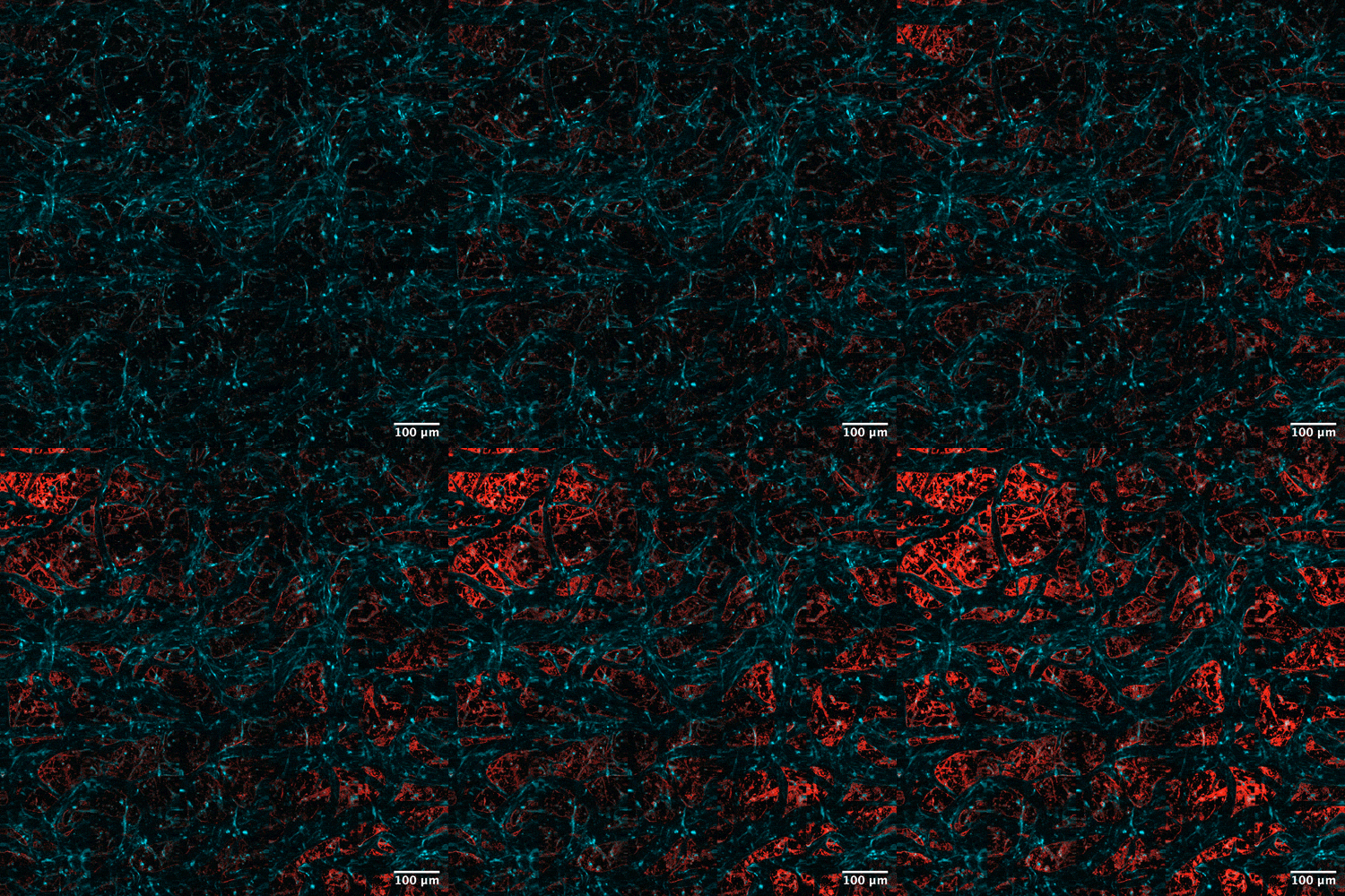
Image: Courtesy of the researchers
The discovery began with experiments led by graduate student Honghao Cao, who pushed a laser system close to its physical limits. As the power approached the fiber’s damage threshold, the light suddenly collapsed into what researchers describe as a “pencil beam,” a tightly confined and stable output.
According to senior author Sixian You, this behavior defies conventional expectations. Instead of becoming chaotic, the light reorganizes itself due to nonlinear interactions within the glass of the fiber. These interactions counterbalance the natural disorder, producing a clean and focused beam without the need for complex optics.
To achieve this effect, the setup must meet two precise conditions. The laser must enter the fiber at an exact angle, and the power must reach a level where nonlinear optical effects become dominant. At that point, the system stabilizes into a highly concentrated beam that maintains its shape.

The implications of this discovery are already being realized in biomedical imaging. The team applied the new beam to study the blood brain barrier, a critical protective layer that controls what enters the brain. Imaging this barrier has traditionally been difficult, with conventional techniques capturing only limited two dimensional views.
Using the new method, researchers were able to generate detailed three dimensional images at cellular resolution and at speeds up to 25 times faster than existing approaches. The beam also eliminates unwanted visual artifacts known as sidelobes, resulting in clearer and more precise images.
Another major advantage is that the technique does not require fluorescent tagging of cells, which is commonly used in biological imaging. This allows scientists to observe natural processes in real time, including how drugs move through the blood brain barrier and interact with specific cell types.
Roger Kamm highlighted the significance of this capability, noting that it could fundamentally change how researchers study drug delivery in the brain. One of the biggest challenges in treating neurological diseases is determining whether medications can effectively reach their targets.
Conditions such as Alzheimer’s disease and Amyotrophic lateral sclerosis have proven particularly difficult to treat in part because of this barrier. The new imaging approach could provide a more accurate way to evaluate drug candidates, especially in human based tissue models.
Looking ahead, the research team plans to further explore the physics behind this self organizing behavior and expand the technique to image neurons directly. There is also interest in commercializing the technology, which could bring advanced imaging capabilities to a wider range of laboratories and medical applications.
This discovery not only reshapes understanding of laser physics but also opens the door to faster, clearer, and more accessible tools for studying the brain, marking an important step forward in both science and medicine.